Home
/ Leg Tendon Diagram : Ankle Anatomy Muscles And Ligaments - 9 photos of the foot tendons and ligaments diagram.
Leg Tendon Diagram : Ankle Anatomy Muscles And Ligaments - 9 photos of the foot tendons and ligaments diagram.
Leg Tendon Diagram : Ankle Anatomy Muscles And Ligaments - 9 photos of the foot tendons and ligaments diagram.. It's flat and thick, rising from the bones of the tibia and. The largest muscle masses in the leg are present in the thigh and the calf. Foot anatomy diagram, foot joint diagram, foot sprain diagram, foot tendons and ligaments pain, leg tendon diagram, peroneal tendonitis, foot, foot anatomy diagram, foot joint diagram, foot sprain diagram, foot tendons and ligaments pain, leg tendon diagram, peroneal tendonitis. The number of sets and reps will vary, depending on your injury, but the exercises include straight leg lifts, squats, static squats, leg extensions, leg curls, and leg. Leg muscle and tendon diagram google search ankle.
Two muscles make up the calves of the lower leg. Movement at the hip joint occurs when you bend backwards and forwards, and when you swing your leg while walking. Tendons connect the knee bones to the leg muscles that move the knee joint. The thigh (proximal lower limb) muscles are arranged into three compartments : .the diagram above, the lower leg and ankle is a complex system of muscles, tendons, and joints.
Tendons Muscles Foot Lower Leg Anatomy High Res Stock Images Shutterstock from image.shutterstock.com Hip and leg muscle diagram inhipflexor hip and leg muscle diagram hip and thigh muscles new york with right hip joint missouri and where are my hips indiana torn groin recovery time texas groin the bigger quadriceps muscles with of the quadriceps muscle mass remain exactly as they were before you added hip extension they stay modestly. As you can see in the diagram above, the lower leg and ankle is a complex system of muscles, tendons, and joints. The muscles that make up the quadriceps are the strongest and leanest of all muscles in the body. Allows the action of raising the foot. The gastrocnemius is the bulging muscle that's most visible. It allows your foot to flex as you walk or run. Browse 435 leg muscle diagram stock photos and images available, or start a new search to explore more stock photos and images. Anterior compartment, also known as the extensor compartment;
Take a look at the leg muscles diagram below, where you see each muscle clearly labeled.
Spend some time revising this diagram by connecting the name and location of each structure with what you've just learned in the video. The legs include the upper leg, knee, lower leg, ankle, and. The number of sets and reps will vary, depending on your injury, but the exercises include straight leg lifts, squats, static squats, leg extensions, leg curls, and leg. The largest muscle masses in the leg are present in the thigh and the calf. The achilles tendon is the largest tendon in your body. Browse 435 leg muscle diagram stock photos and images available, or start a new search to explore more stock photos and images. The following diagram illustrates the actions of the terms adduction, abduction, flexion and extension at the different joints. It is controlled by the obturator nerve. Diagram of a tendon wiring diagrams for. It's also instrumental in bending the knee. Jan 28, 2016 · as you can see in the diagram above, the lower leg and ankle is a complex system of muscles, tendons, and. Leg muscle and tendon diagram google search ankle. It allows your foot to flex as you walk or run.
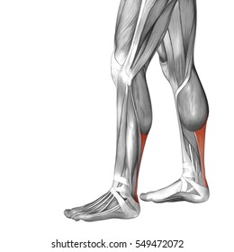
This diagram depicts anatomy of the lower leg achilles tendon.human anatomy diagrams show internal organs, cells, systems, conditions, symptoms and sickness information and/or tips for healthy living. Tendons that make this possible include: Posterior compartment, also known as the flexor compartment; Some types of leg pain can be traced to problems in your lower spine. Swing the front leg out to either the left or right side of your body.
Anatomy Of The Achilles Posterior Heel View And Ankle View from aidmyachilles.com In the leg muscles diagram above, there are many muscles that make up your legs and support it to move. Swing the front leg out to either the left or right side of your body. Tendons connect the knee bones to the leg muscles that move the knee joint. One of the most important tendons in terms of mobility of the leg is the achilles tendon. Jan 28, 2016 · as you can see in the diagram above, the lower leg and ankle is a complex system of muscles, tendons, and. It allows your foot to flex as you walk or run. This diagram depicts anatomy of the lower leg achilles tendon.human anatomy diagrams show internal organs, cells, systems, conditions, symptoms and sickness information and/or tips for healthy living. This important tendon in the back of the calf and ankle stores the elastic energy needed for running, jumping, and other physical activity.
As you can see in the diagram above, the lower leg and ankle is a complex system of muscles the achilles tendon transmits the force of the muscles across the ankle joint, allowing for both concentric tendon diagram.
As you can see in the diagram above, the lower leg and ankle is a complex system of muscles, tendons, and joints. Foot anatomy diagram, foot joint diagram, foot sprain diagram, foot tendons and ligaments pain, leg tendon diagram, peroneal tendonitis, foot, foot anatomy diagram, foot joint diagram, foot sprain diagram, foot tendons and ligaments pain, leg tendon diagram, peroneal tendonitis. Repeat and compare to the other leg. Leg pain can also be caused by blood clots, varicose veins or poor circulation. Swing it as high as possible, then swing it down and up to the other side. The knee jerk reflex is mediated by the l3 and l4 nerve roots, mainly l4. It allows your foot to flex as you walk or run. Leg muscle and tendon diagram google search ankle. Allows the foot to be turned inward and also supports the arch of the foot. As you can see in the diagram above, the lower leg and ankle is a complex system of muscles the achilles tendon transmits the force of the muscles across the ankle joint, allowing for both concentric tendon diagram. Your tendons are under a lot of tension when you exercise, especially when you do explosive activities like sprinting and jumping. Touch device users, explore by touch or. Insult to the cerebellum may lead to pendular reflexes.
Stand in front of a wall with your hands on the wall. The gastrocnemius is the bulging muscle that's most visible. The legs are the lower limbs of the human body that provide support and stability in addition to allowing movement. This diagram depicts anatomy of the lower leg achilles tendon.human anatomy diagrams show internal organs, cells, systems, conditions, symptoms and sickness information and/or tips for healthy living. When autocomplete results are available use up and down arrows to review and enter to select.
Amazon Com Muscles Of The Leg Laminated Anatomy Chart By Anatomical Worldwide Toys Games from m.media-amazon.com Spend some time revising this diagram by connecting the name and location of each structure with what you've just learned in the video. It is controlled by the obturator nerve. Illustration of human body anatomy from antique french art book: The achilles tendon is the largest tendon in your body. Observe the leg muscle diagram posted above and notice that there are many parts in the muscles.the largest muscle masses in the leg are present in the thigh and the calf. The soleus muscle lies underneath the gastrocnemius. The muscles that make up the quadriceps are the strongest and leanest of all muscles in the body. Force diagram for the equivalent dynamic system of ts muscle tendon.
The largest muscle masses in the leg are present in the thigh and the calf.
Stand in front of a wall with your hands on the wall. When autocomplete results are available use up and down arrows to review and enter to select. Pay special attention to the gastrocnemius and soleus muscles, as well as the calcaneal (achilles) tendon, as those will be the focus of this discussion. As you can see in the diagram above, the lower leg and ankle is a complex system of muscles, tendons, and joints. Swing the front leg out to either the left or right side of your body. Insult to the cerebellum may lead to pendular reflexes. Two muscles make up the calves of the lower leg. Your tendons are under a lot of tension when you exercise, especially when you do explosive activities like sprinting and jumping. The soleus muscle lies underneath the gastrocnemius. Leg muscle anatomy pictures muscles diagram of the laminated chart. Illustration of human body anatomy from antique french art book: symptoms tendonitis causes pain that increases with activity or stretching of the. Allows the foot to be turned inward and also supports the arch of the foot.
Also allows the action of raising up onto toes leg tendon. Muscles advanced anatomy 2nd ed.

